
Magnification Dentistry
Magnification Dentistry
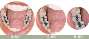 Is magnification dentistry for you? As technology continues to advance, the field of dentistry is no exception for undergoing innovation. Magnification is the current standard of dental care, yet only a small percentage of dentists take advantage of this technology. Both dental professionals and patient benefit from the ability to magnify an image 2–4.5x with standard loupes. This ability is an asset, which allows dentists increased visibility to diagnose and provide treatment for a patient. Let’s take a look at some of the benefits of using microscopes in dentistry.
A loupe is a small magnification device that is typically used by dentists as an attachment to glasses. While loupes do provide a closer look, a dental microscope allows for a range of magnification from 2.6-16x. The average microscope used by a dentist operates at an 8x magnification as opposed to the 2.5-4.5x magnification provided by a loupe. With greater magnification comes an improved accuracy and subsequent treatment. Dentists can accurately prepare and deliver a more conservative treatment, which is beneficial to the surrounding teeth and tissues. Ultimately, a higher magnification provides higher visibility in all areas of dentistry from diagnosing to prepping, seating and finishing restorations.
As a dentist, you’re constantly bending over and putting your body into uncomfortable position. By using a scope, ergonomics will be improved. Dentists are essentially forced to sit up straight with proper posture to use the tool. This bettered posture will reduce neck and back strain, leaving you feeling more refreshed at the end of the day.
Along with a more comfortable body position, dentists who use a microscope can also reduce their eye fatigue. Many dentists who use loupes often feel the strain of the converging vision caused by using a loupe. A microscope allows for a higher magnification, which opens up the parallel vision of the working field, reducing both strain and fatigue.
The built-in lighting on modern microscopes provides incredible visibility to even the most difficult areas of a patient’s mouth. The xenon, LED, or halogen lights can reach spaces, giving dentists more visibility in their working field and the ability to discover any hidden decay or other issues. For example, a deep interproximal decay on the mesial of second molars can be found by the light, increased visibility when looking down into a post space, or when completing a composite restoration.
Finally, a microscope can also be paired with a camera and monitor, so that dental procedures can be documented. These recordings can be used to help educate and communicate with patients and other dentists. Additionally, they can help your staff learn more about the procedures they are performing or assisting with.
It’s important to note that there are different challenges many dentists face when implementing microscopes into a clinic such as staff acceptance, the cost difference, the learning curve, and longer procedures. Despite these inconveniences, the benefits are incredible. Many dentists find that it’s hard to go back to a loupe after using a microscope.